Laser Scanning Microscope Max Magnification

What Is Confocal Laser Scanning Microscopy

Confocal Laser Scanning Microscopy An Overview Sciencedirect Topics
Confocal Microscopy Introduction Olympus Life Science
Confocal Microscopy Confocal Microscope Scanning Systems Olympus Life Science
Fv3000 Confocal Laser Scanning Microscope From Olympus Life Science Solutions Get Quote Rfq Price Or Buy

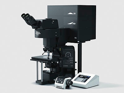
Inverted Zeiss Lsm880 Laser Scanning Confocal Microscope With Airyscan Cell Sciences Imaging Facility Csif

Pongali raghavendra thammineni pullaiah in advances in cell and molecular diagnostics 2018.
Laser scanning microscope max magnification.

Confocal Laser Scanning Microscope Labcompare Com
Confocal Microscopy Resolution And Contrast In Confocal Microscopy Olympus Life Science
Micro Small Scopeo To Watch Microscopy The Relative Sizes Of Molecules Cells And Organisms Ppt Download

A Practical Guide For Fluorescent Confocal Microscopy The Marder Lab

Zeiss Lsm 900 Confocal Laser Scanning Microscope From Carl Zeiss Microscopy Biocompare Com

Laser Scanning Microscope 13 Steps With Pictures Instructables

Overview Of Confocal Laser Scanning Microscopes

Advantages And Limitations Of Scanning Electron Microscopy Sem And Download Table

Zeiss Microscopy Online Campus Introduction To Spinning Disk Microscopy

Olympus News Release Confocal Laser Scanning Microscope Ols 3000 World S Highest Resolution Of 0 12µm

Confocal Laser Scanning Microscope An Overview Sciencedirect Topics

33 Laser Scanning Confocal Microscopy And Laser Microdissection Musculoskeletal Key

Laser Scanning Microscopes Keyence America

Rp Photonics Encyclopedia Optical Profilometers Non Contact Optical Surface Profile Measurements Interferometer Coherence Oct Focus Variation Digital Holography Triangulation Time Of Flight Structured Light Performance Factors Geometric
Olympus Launches Two Types Of Upright Fv3000 Confocal Laser Scanning Microscope 2017 News Olympus
Scanning Optical Microscopy Emmi Laser Scan Obirch Som

Laser Scanning An Overview Sciencedirect Topics
Confocal Microscopy Confocal Microscope Objectives Olympus Life Science
Https Encrypted Tbn0 Gstatic Com Images Q Tbn 3aand9gcr3fysoxor5w4y0kayjtt5nby84 Yhi3vdxn3rx2 E Usqp Cau

Adaptive Optics For Biomedical Microscopy Optics Photonics News
.jpg?rev=33A9)
Fv1200 Olympus Life Science

Inverted Zeiss Lsm 780 Multiphoton Laser Scanning Confocal Microscope Cell Sciences Imaging Facility Csif

Imaging Surface Characterisation Australian National Fabrication Facility Queensland Node

Confocal Laser Scanning Microscope Specification Price Image Bio Equip In China
Source : pinterest.com
