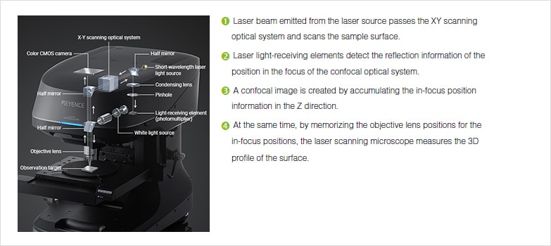
Laser Scanning Confocal Microscope Magnification And Resolution

What Is Confocal Laser Scanning Microscopy

Confocal Laser Scanning Microscopy An Overview Sciencedirect Topics
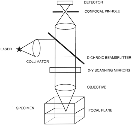
How Does A Confocal Microscope Work
Confocal Laser Scanning Microscopy Creative Biolabs
Confocal Microscopy Introduction Olympus Life Science
Confocal Microscopy Confocal Microscope Scanning Systems Olympus Life Science

Embryos and construct 3 d structures from the obtained images.
Laser scanning confocal microscope magnification and resolution.
Profile Measuring Laser Microscopes Instruments Used For Roughness Measurements Introduction To Roughness Keyence America

New Microscope Shows The Quantum World In Crazy Detail Quantum World Electron Microscope Properties Of Materials

33 Laser Scanning Confocal Microscopy And Laser Microdissection Musculoskeletal Key

Winners Of The 2019 Nikon Small World Photomicrography Competition Show The Artistry Of Science In 2020 Nikon Small World Microscopic Photography Best Microscope
A Robust And Versatile Platform For Image Scanning Microscopy Enabling Super Resolution Flim Nature Methods

Confocal Laser Scanning Microscope An Overview Sciencedirect Topics

How The Confocal Laser Scanning Microscope Entered Biological Research Sciencedirect

Amazing Images The Best Science Photos Of The Week Things Under A Microscope Microscopic Photography Confocal Microscopy
Zemax Optical Model Of A Simplified Scanning Microscope The Rays Are Download Scientific Diagram
Confocal Microscopes Microscope Imaging Network

Moth Antennae Detail Microscopic Photography Insect Photos

Confocal Laser Scanning Microscopy An Overview Sciencedirect Topics
Confocal Laser Scanning Microscopy Springerlink

Invisible Details Of Tiny Creatures Uncovered With Laser Microscope Photos Microscopic Photography Macro Photography Insect Photos
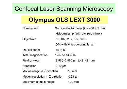
Confocal Laser Scanning Microscopy Olympus Ols Lext 3000
Confocal Microscopy Of The Eye
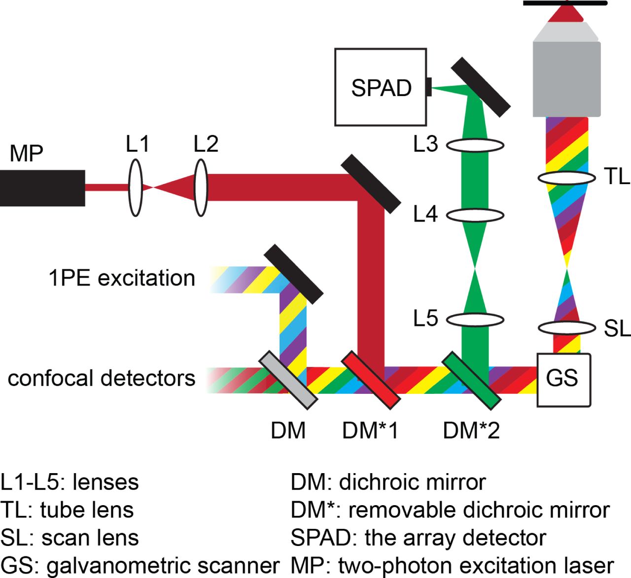
Easy Two Photon Image Scanning Microscopy With Spad Array And Blind Image Reconstruction Biorxiv

Pin By Lucy Connell On Sem Revelations In 2020 Scanning Electron Micrograph Electrons Stock Photos
Https Encrypted Tbn0 Gstatic Com Images Q Tbn 3aand9gcr3fysoxor5w4y0kayjtt5nby84 Yhi3vdxn3rx2 E Usqp Cau
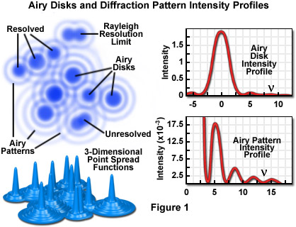
Confocal Microscopy Resolution And Contrast In Confocal Microscopy Olympus Life Science

Problems Solved

Wistar Rat Retina Nikon Small World Micro Photography Small World

Asa De Borboleta Microscopic Photography Macro Photography Micro Photography

Confocal Microscopy An Overview Sciencedirect Topics
Source : pinterest.com
